
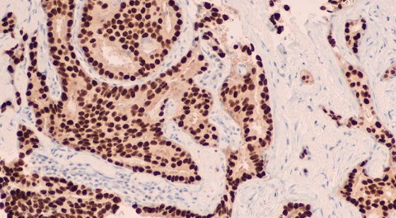
The immunohistochemistry (IHC) is a technique commonly applied in the field of both research and clinical diagnosis. The use of antibodies on a section of tissue allows the specific proteins of interest to be detected, to later determine both their location and abundance by microscopy.
Although, in principle, the protocol may seem relatively simple to apply, it requires optimizing some parameters that will be essential for the good resolution of each test. The same protocol can work perfectly with one antibody, but not with others; and some antibodies may need specific adjustments in some of the steps of the procedure.
In this entry we collect some key considerations to optimize the results and solve problems in Immunohistochemistry (IHC) .
How To Troubleshoot Immunohistochemistry (Ihc)
Below we detail the possible causes (PC) that can lead to ambiguous or erroneous results, as well as the tips or solutions (S) to solve them:
1.- Absence Of Stain
– Antigens –
PC: Absence of antigen, or presence in very low quantity
- S1: Analyze protein expression by in situ hybridization.
- S2: Include an amplification step of the detection signal in the protocol.
- S3: Increase the concentration of the antibody.
PC: Alteration of epitopes during the fixation step
- S: Try to restore immunoreactivity using antigen recovery techniques.
PC: Ineffective antigen recovery
- S1: Increase the treatment time.
- S2: Change the recovery technique.
PC: The protein is located in the nucleus and the antibody cannot penetrate
- S: Add permeabilizing agents to the blocking buffer and to the antibody dilution buffer.
– Antibodies –
PC: Antibodies have lost activity
- S1: Follow manufacturer’s instructions regarding antibody storage criteria. (You can find some tips for properly storing antibodies in this post .)
- S2: Always include positive controls to rule out antibody malfunction.
PC: Incompatibility between primary and secondary antibody
- S: The secondary antibody must be directed against the species in which the primary antibody was generated. ( Here you can consult a Guide to select secondary antibodies .)
PC: Incorrect primary antibody
- S: Select a specific antibody against the antigen of interest. It should be borne in mind that in immunohistochemistry, the antibody must recognize the native conformation of the antigen, so it is worth contrasting in the technical sheet that has been validated for use in this application.
– Sample preparation –
PC: Inadequate tissue fixation
- S1: Increase the fixing time.
- S2: Try a different fixer.
PC: Over-fixation of the tissue
- S: Reduce the duration of the dive or the post-fixation steps.
– Reagents –
PC: Reagents have been added in an incorrect order and / or steps have been omitted
- S: Carefully review the procedure that has been carried out.
2.- Weak Stain Of Protein Diana
– Antigens –
PC: Inadequate antigen recovery
- S1: Vary the recovery conditions.
- S2: Change the recovery method
PC: Alteration of the electrostatic charge of the antigen
- S: Adjust the pH or cationic concentration of the antibody buffer.
– Antibodies –
PC: Low reactivity of the primary antibody
- S1: Ensure that the pH of the antibody diluent is within the optimal range specified for antibody binding (pH 7-8).
- S2: Make sure that the antibody has been stored according to the manufacturer’s instructions.
- S3: Increase the concentration of the primary antibody and / or the incubation time.
PC: Inhibition of secondary antibody
- S1: Reduce the concentration of the secondary antibody. (While high concentrations of secondary antibody may increase background staining, extremely high concentrations may have the opposite effect, reducing antigen detection.)
- S2: If the diluent contains antibodies that neutralize the antigen, these will block the binding of the secondary antibody. Remove neutralizing antibodies or change diluent.
– Sample preparation –
PC: Improper fixing method
- S1: Increase the fixing time.
- S2: Try a different fixing method.
– Reagents –
PC: enzyme – substrate reactivity
- S1: Change the enzyme diluent.
- S2: Prepare the substrate again at a suitable pH.
3.- High Background Noise
– Antibodies –
PC: Secondary antibody exhibits cross reactivity or nonspecific binding
- S1: Treat the tissue with normal serum of the species that the secondary antibody.
- S2: Use pre-adsorbed antibody against the species of the sample.
PC: Non-specificity of the primary antibody
- S1: Reduce the concentration of the primary antibody
- S2: Increase the concentration of the blocking buffer and reduce the time between blocking and the addition of the primary antibody.
- S3: Use a different primary antibody.
PC: High concentration of primary or secondary antibody
- S: Titrate the antibody to determine the optimal working concentration.
PC: Hydrophobic interactions between the antibody and other proteins present in the tissue
- S: Reduce the ionic strength of the antibody diluent.
PC: incubation time or temperature too high
- S: Reduce the incubation time and / or temperature.
PC: Inadequate washing of sections
- S: Wash at least three times between each of the steps of the procedure.
– Sample preparation –
PC: Presence in the tissue of endogenous enzymes (peroxidases and / or phosphatases)
- S1: Block peroxidases with hydrogen peroxide in methanol before incubation with the primary antibody.
- S2: Inhibit the action of endogenous phosphatases with levamisole.
PC: Presence of endogenous biotin
- S: Block endogenous biotin activity by avidin / biotin blocking reagent before incubation with the primary antibody.
PC: The fabric sections have dried
- S: Prevent the tissue from drying out during the staining process.
The Immunohistochemistry (IHC) technique, like other immunoassays, involves various steps that cannot be universally optimized, having to adjust certain conditions for each of the assays, which translates into a multitude of variables that can affect the the results are as expected.